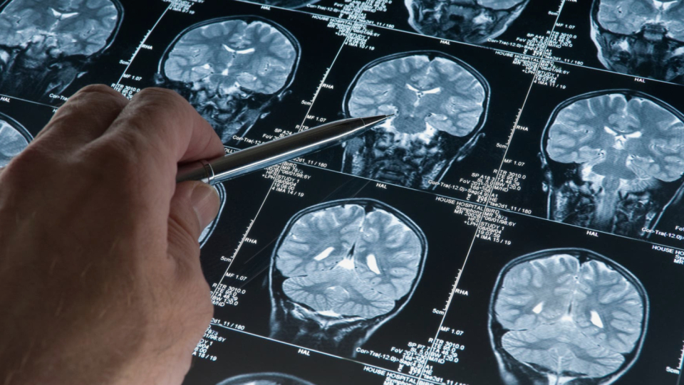
The neuroscientist Jakob Seidlitz left the practice dissatisfied after he had presented his 15-year-old son there to the pediatrician for a check-up. There was no problem with his son – he seemed to develop normally for his age, according to the height and weight tables that the medic used. But what was missing, according to Seidlitz's feelings, was a comparable yardstick to evaluate the development of the brain. "It is shocking how little biological information medicine has about this critical organ," says the neurologist from the University of Pennsylvania.
He could change that soon. Together with colleagues, he has accumulated more than 120,000 brain scans, the largest collection of this kind. With them, he wants to create the first comprehensive growth tickets for the brain. The illustrations show how the brain grows quickly at a young age and then slowly shrinks again in old age. The sheer size of the study published on April 6th in "Nature" has amazed the specialist community, which for a long time had problems with the quality of its data - among other things, because the samples are often very small. Core resonance imaging is expensive, which is why research groups can often only recruit a small number of test subjects.
"The massive data set they've assembled is extraordinarily impressive and sets standards for the whole field," says Angela Laird, a neuroscientist at Florida International University in Miami.
Several solutions because of the pandemic
Nevertheless, the authors point out that their database is not complete – they struggled to collect brain scans from all regions of the world. The resulting diagrams are therefore only a first draft, which still needs to be revised for use in clinical practice.
When the diagrams are finally passed on to pediatricians, care must be taken to ensure that they are not misinterpreted, says Hannah Tully, a pediatric neurologist at the University of Washington in Seattle: "A big brain is not necessarily a well-functioning brain.«
Since the brain structure is very different from person to person, the researchers had to summarize a large number of scans to get statistically significant data for a meaningful series of growth diagrams. This is not an easy task, says Richard Bethlehem, neuroscientist at the University of Cambridge and co -author of the study. Instead of even carrying out thousands of scans, which would take decades and would be unaffordable, the researchers used the neuroimaging studies that have already been completed.
Bethlehem and Seidlitz sent emails to researchers around the world, asking if they would provide their neuroimaging data for the project. The two were amazed at the number of responses, which they attribute to the fact that the Covid-19 pandemic left researchers less time in their labs and more time than usual for their email inboxes.
The team collected a total of 123 894 scans of 101,457 people who ranged from the fetus 16 weeks after conception to 100-year-old adults. The scans included brains of neurotypical people and people with a variety of diseases such as Alzheimer's .. The researchers used statistical models to extract information from the images and ensure that the scans were directly comparable - regardless of the type of MRI -was used.
A diplomatic masterpiece
The end result is a series of graphs that record various important brain metrics by age. Some measures, such as the volume of gray matter and the average cortical thickness – the width of gray matter, reach their peak in the early period of a person's development, while the volume of white matter, which is located in the depth of the brain, reaches its peak around the age of 30. Especially the data on the volume of the ventricles (the amount of cerebrospinal fluid in the brain) surprised Bethlehem and Seidlitz. Although scientists knew that this volume increases with age, as it is usually associated with brain atrophy, but Bethlehem was shocked by how quickly it increases in late adulthood.
The study follows a groundbreaking work, published on March 16 in "Nature", which shows that most experiments with imaging methods of the brain contain too few scans. As a result, you cannot reliably recognize relationships between brain function and behavior, which means that your conclusions could be wrong. In view of this result, Laird expects science to turn to similar concepts as that of Seidlitz and Bethlehem to increase statistical meaning.
Compiling so many data sets is a "diplomatic masterpiece," says Nico Dosenbach, a neuroscientist at Washington University in St. Louis who co-authored the March 16 study. In his opinion, this is the standard by which researchers should orient themselves when compiling brain images.
Despite the size of the data record, Seidlitz, Bethlehem and her colleagues admit that their study suffers from a problem that is typical of neuroimaging studies-a remarkable lack of diversity. The brain scans collected by them come mainly from North America and Europe and reflect disproportionately many white, urban and wealthy population groups in university age. This limits the generalizability of the results, says Sarah-Jayne Blakemore, a scientist for cognitive neurosciences at the University of Cambridge. The study includes only three data records from South America and one from Africa - this corresponds to about one percent of all brain scans used in the study.
According to Laird, billions of people worldwide do not have access to MRI machines, making it difficult to access diverse brain image data. But the authors haven't stopped trying. They have set up a website where they want to update their growth charts in real time as soon as they receive further brain scans.
Open science is not compensated
Another challenge was to appreciate the origin of the brain scans used for the creation of the diagrams. Some of the scans came from open access data records, others were not freely accessible. Most scans in such closed data records had not yet been processed so that they could be integrated into the growth diagrams. This service then also provided the experts who belong to the data records. They are therefore listed among the authors of publication.
The owners of the open datasets, on the other hand, were only quoted in the paper – which doesn't mean as much prestige for researchers seeking funding, collaborations, and promotions. Seidlitz, Bethlehem and their colleagues processed this data. In most cases, Bethlehem said, there was virtually no direct contact with the owners of these records. The paper lists about 200 authors and cites the work of hundreds of others who have contributed brain scans.
There are a number of reasons why records are closed: for example, to protect the privacy of health data or because the working groups involved do not have the resources to publish them. But that doesn't make it fair that the researchers who opened their datasets didn't receive authorship, the study's authors say. As they write in the supplementary information to their work, the situation "perversely constitutes an incentive against open science, since the people who do the most to make their data openly accessible are the least likely to deserve recognition".
Ultimately, these concerns can be attributed to how researchers are evaluated by science, says Kaja Lewinn, a social epidemiologist at the University of California in San Francisco, who deals with neuro development. According to her, it is the task of all those involved - including the donors, magazines and research institutions - to re -evaluate how brain research can be appropriately recognized and rewarded, especially since these types of large -scale studies are increasingly carried out.
© Springer Nature 10.1038/d41586-022-00971-1, 2022


