


Scientific knowledge can arise in the strangest moments - for example when a rat in the laboratory makes the "looking down dog". This yoga exercise shows that the rodents benefit from the associated stretches like humans. Researchers are increasingly revealing the importance of a tissue that science has overlooked for centuries.
What we know today as fascia, the anatomist Erasmus Wilson called natural bandages in the 19th century. Exposed, they look exactly like this: long strips of white, fibrous connective tissue. Fascia is strong yet flexible. This makes them perfect for keeping muscles and organs in place. But they are also sticky, mushy and obstruct the view of the muscles, bones and organs that cover them. This explains why anatomists cut off this tissue for years, threw it away, and gave it little thought.
Recently, however, the view of the fascia has begun to change. Researchers have found that they are anything but a passive insulation layer. Rather, they are a place of biological activity that could explain some of the links between lifestyle and health. It may even be a kind of sensory organ. "There's a lot more going on in the fascia than you might think," says Karl Lewis of Cornell University in Ithaca, New York.
Experts have now recognized that a better understanding of this ubiquitous tissue is urgently needed. It could open up new ways to combat widespread but difficult to treat diseases - from diseases of the immune system to chronic pain.
What holds the body together
Examining fascia is difficult, among other things, because there is disagreement about what they actually are. They fall under the term connective tissue. This includes not only tendons and ligaments, but also in the broadest sense, bones, skin and adipose tissue.
However, most fascia researchers understand this to mean tissue layers made of strong collagen and more stretchable elastin fibers. These layers are separated in many places by looser, so-called areolar fasciae. They consist of a few fibers, and their interstices are filled with a slimy substance. This allows adjacent layers to slide on top of each other. The main components of the lubricant are hyaluronic acid and proteoglycans. While the former have an attractive and thus moisturizing effect on water, the latter provide good cushioning. Both the fascial fibers and the lube are formed by certain cells in the tissue: the fibroblasts and the recently discovered fasciocytes.
If you were cutting open the body lengthways to make your natural shell visible, you would find two clearly delimited layers: the superficial fascia directly under the skin as well as the deep fascia, the muscles and organs envelop. Some experts also include the visceral fascia, which lines the body cavity and divides into the rooms of the various organs. There are also thin layers of connective tissue that cover pretty much every inner surface of the body. According to this definition, the fascia form a network that holds people together (see "a body -wide network").
Surprisingly, until the early 2000s, no one looked at the fabric in detail. Carla Stecco was among the first to do this. She is an orthopedic surgeon and anatomist at the University of Padua in Italy. Already 20 years ago she began to study the fascia. The occasion: Her father, the physiotherapist Luigi Stecco, invented a form of physical therapy, which he called fascia manipulation. So, he claimed, you can treat everything from headaches to muscle and joint pain. Today, his approach is one of many physical therapies based on the idea that fascia can be stiffened and loosened by massage.
A body -wide network
Our understanding of how fascia affects health depends on how to define the beginning and end of the fascia in the body.
Some people believe that the term not only includes the different layers of the connective tissue under the skin and around the muscles, but also the interstitium. This is that fluid -filled connective tissue that lines each organ, every muscle fiber and every blood vessel.
If this is true, the fasciae form a fluid-filled network: this runs through the entire body and could act both as a shock absorber and as its own immune system, which plays a role in inflammatory diseases, scarring and the spread of cancer.
The true nature of the interstitium was only discovered in 2018. In a study, Neil Theise from the Icahn School of Medicine in Mount Sinai, New York, and his colleagues applied a new microscopic technology to examine the structure of the interstitium on the living person. In the past, the tissue could only be viewed by taking it out and crushing it on an object carrier. When looking at the tissue, it was shown that what has so far looked like a dense tangle of fibers has a spongy structure. It is filled with liquid that flows into the lymphatic system and is therefore part of the body's immune system.
The team suspects that physical exercise can help to keep this fluid system healthy: through the higher pumping capacity of the heart, the mobilization of the digestive tract and the movement of the body. "Apparently, such spaces are not static," says Theise. That discovery suggests that the body is interconnected in a way that we are only just beginning to understand.
The problem with this: There was no evidence of or against the thesis that touch and grabs do something with the fascia or even affect the pain. Carla Stecco found that the literature did not explain in detail what fascia actually are. It was not even known whether they were linked to nerves, she says.
Meanwhile, she and others have shown that fasciae are abundantly endowed with nerve fibers. The information they pass on varies depending on the part of the body. The nerves of the superficial fascia are specialized for pressure, temperature and movement. The deep fascia are involved in proprioception, that is, they establish where the body is located in space. In addition, they can perceive pain, specialists speak of nociception.
The fascia - your own organ?
In view of the sensory function, some scientists believe that the fascia should be considered as a separate organ that has the task of transmitting messages from the inside of the body. Robert Schleip from the Technical University of Munich recently estimated that the fascia of an adult contains about 250 million nerve endings – a similar number or even more than the skin. "They are undoubtedly our most nervous sensory organ," he says. Others are a little more cautious. "There is a strict definition of what an organ is. This is about the material organization, the cell types and the function. The fascia is a hot candidate," says Lewis. "But it is still too early to make this decision".
Organ or not, there is indications that deep fascia send a different kind of messages than other body tissue. Experiments in which healthy test subjects voluntarily be given painful injections in skin, muscles or fascia have shown that the nerves in skin and muscles react with concentrated, locally limited pain signals. The nerve network in the fascia, on the other hand, called out a large, difficult pain. Such diffuse pain is a characteristic of different chronic pain diseases, for example fibromyalgia.
Some study authors have associated the disease with inflammation of the fascia. Muscle soreness has also been attributed to damage to the muscles for a long time. In the meantime, however, some researchers assume that this has more to do with injuries or inflammation in the fascia.
The bad news for people with inflamed fascia: if this condition lasts for a long time, the composition of the nerves located there changes, they become more sensitive to pain. In rats, the proportion of nociceptive fibers – those that are equipped with pain receptors – increased from four to 15 percent after chronic inflammation of the deep fascia in the lower back. That might explain why low back pain is so difficult to treat. They are one of the most common causes of work stoppages and general movement restrictions. 85 percent of cases worldwide are considered non-specific, which means that the exact cause cannot be determined.
The thoracolumbar fascia could be a good place to look for the cause of back pain. This is a diamond-shaped, multi-layered structure in the lower back, the layers of which are connected to various muscle groups in the trunk. "The thoracolumbar fascia is like a big sensor. He feels the tension coming from the upper limbs, the spine and the abdomen," says Stecco. Perhaps the sensory neurons in the fascia respond to this tension by registering it as pain.
In addition to changes in the nerves, inflammation in the loose, areolary fascia can make the situation worse. Using ultrasound images of the lower back, Helene Langevin from the US National Institute of Health in Maryland has shown that the thoracolumbale fascia is 20 percent stiffer in people with chronic back pain than in pain-free. Several, glued tissue layers are the cause, it said.
Langevin's studies on pigs confirmed this thesis. The team also showed how a lack of movement in the lower back makes the fascia stiff and causes adhesions in places where two layers joined together with the help of new collagen fibers – even if an initial injury had already healed. According to other studies, this restricts movement. And not only in the fascia surrounding the stiff spot, but also in neighboring regions. In particularly severe cases, the fascial layers can stick together to form an immovable block that extends from the superficial fascia to the deep fascia and into the muscle.
In addition to injuries and inflammation, there are numerous other reasons why fascia are stiff. The activation of the sympathetic nervous system, which is involved in the body's combat or escape reaction, could get the fascia to contract. Schleip's studies indicate this. The system causes the fibroblasts to convert into myofibroblasts. These cells are crucial for inflammatory reactions as a result of injuries, as well as joint problems such as a stiff shoulder.
How stress leads to such a stiffening has not yet been sufficiently researched. According to Schleip, adrenaline increases the expression of an inflammatory substance called TGF-beta. This is stored in the loose fascia to prepare the body for the next stress. When this happens, "the fibroblasts drink [TGF-beta] and become myofibroblasts within a few hours," says Schleip. That makes them four times as strong. "They are contraction machines.«
Adrenaline is not apparently the only factor that affects the stretching of the fascia. "Estrogen makes them more elastic," says Stecco. »Fascia is a very dynamic tissue that can react to hormonal, chemical and mechanical influences. Everything together determines whether they are elastic or stiff. "
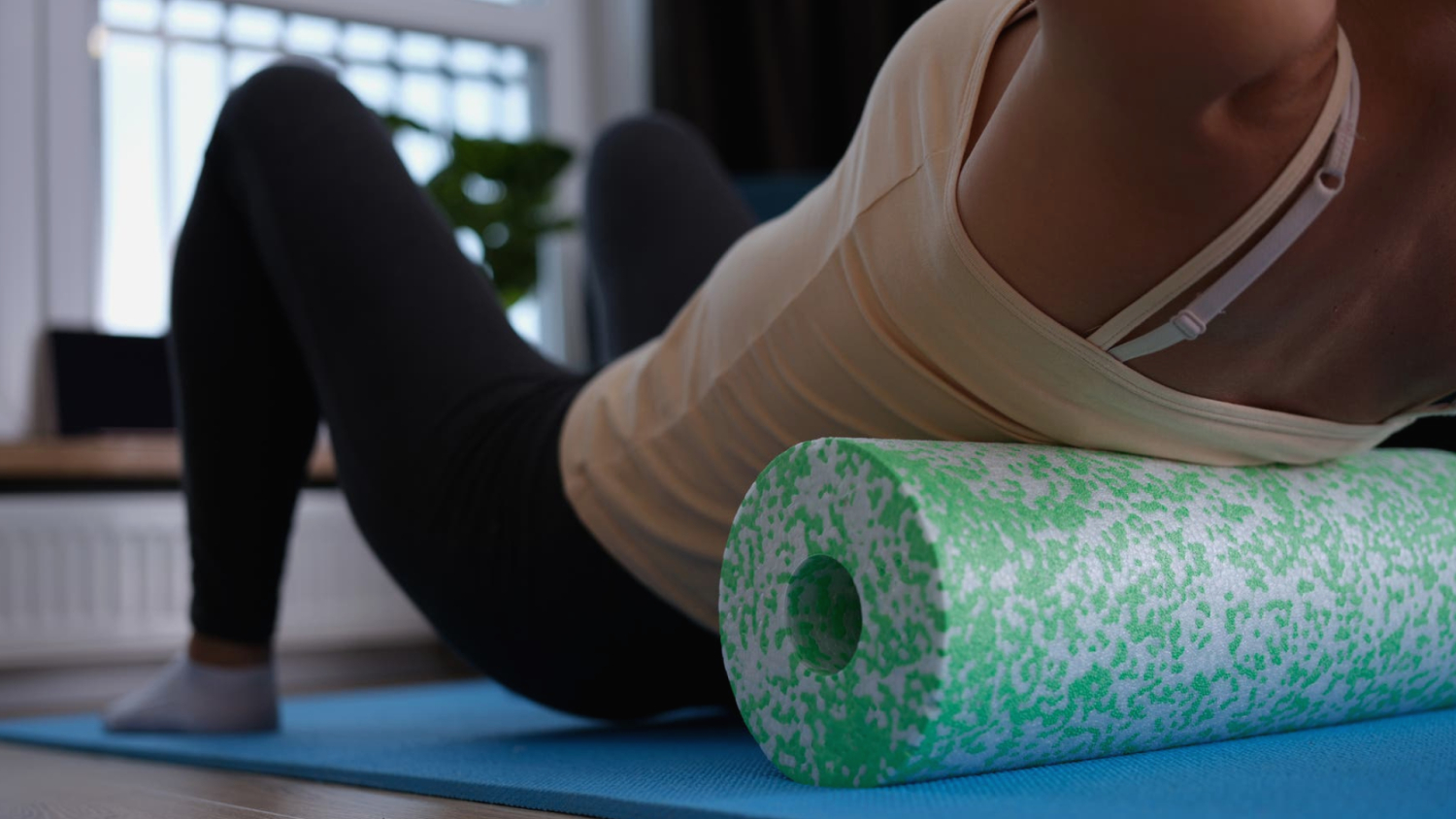
The good thing about the dynamic nature of the fascia: if people change their lifestyle, some problems may be solved. One promising measure that is currently being studied is stretching. In tissue samples from rats, Langevin observed that stretching alters the fibroblasts that form the scaffold of the areolar fibroblasts. They become several times larger, longer and flatter. "The stretching of the tissue allows him to relax, " she adds.
Stretch could help
Stretching the lower back twice a day for five minutes, as Langevin's studies with pigs show, can not only reduce the size of the inflamed area, but should also ensure that anti-inflammatory substances are secreted in the fascia. This is a promising result, because chronic inflammation is associated with almost all ailments – from heart disease to cancer and depression.
A Harvard Medical School team is currently carrying out a study with people to find out whether the results can be transferred. A pilot study completed at the end of 2021 shows that healthy volunteers who completed a one -hour stretching unit showed different levels of certain immune molecules, the cytokines, as the control group that did not stretch. Accordingly, stretching would affect the inflammation.
Future studies should clarify whether the content of resolvins is also increasing. The body produces these substances to curb inflammation. This was the case with rats and pigs. If it's similar in humans, stretching could help reduce widespread chronic inflammation triggered by long-term stress, obesity, and poor diet.
So far, it is unclear whether physical therapies such as massages, which focus on loosening the fascia, have the same cellular and anti-inflammatory effects as stretching. Maybe they only bring about short-term changes. For example, it could be that manual therapies heat the tissue. In this way, the fascial matrix is demonstrably less viscous and the layers can slide more easily temporarily. However, Langevin urges caution: as long as one does not know exactly what happens with such therapies, it is unclear what they do with the fascia or whether they even have any effect.
In order to implement the research results into evidence -based treatments, the tissue must also overcome its image problem. This goes back to the 1940s and 1950s when medical research paid little attention to the fascia. Instead, it became the focus of an alternative health approach that the deceased biochemist Ida Rolf had developed. Her method, which she called structural integration - better known as Rolfing - is a mixture of physical therapy and theories about the orientation of the body's own energy fields. Since then, the fascia have become a "buzzword" in alternative medicine.
According to Stecco, it is high time that conventional medicine pays more attention to fascia. The tissue is important for general health, says Stecco. This would be "the true revolution of fascia".