


»Cartilage does not heal.« That's what doctors often say when the flexible tissue lining our hips, knees and shoulders is injured or worn out so much by osteoarthritis that the joints hurt with every movement. Orthopaedists have also explained to me that cartilage is not supplied with blood, which is why neither repair tools nor nutrients can reach the injured area. I found this only implausible: Why should living tissue not be able to replace damaged cells? And indeed, research suggests that cartilage – at least that in our joints – has a limited ability to repair. This raises hopes for treatments that can heal cartilage or protect already damaged tissue from further degradation.
To imagine what articular cartilage looks like, think of the tough, white layer at the end of a chicken bone. Most of it is a sponge-like material, the so-called extracellular matrix. It consists of water and fibrous proteins and is produced by special cells called chondrocytes. "Every tissue – apart from the enamel – has the ability to regenerate itself," explains rheumatologist Virginia Kraus from Duke University School of Medicine in North Carolina, USA. "New tissue is formed, old tissue is crushed and washed away." However, the renewal process in the cartilage is very sluggish, she says. And it is also true that the tissue is not supplied with blood in adults. Instead, the cartilage receives nutrition through a process that experts call a dynamic load: if we put weight or pressure on a joint, nutrient-containing joint fluid flows in and out. "That's why exercise is so important for joint health," explains Kraus. "As a result, the cartilage is supplied with nutrients.«
Kraus is one of the few scientists who examine how the tissue is slowly renewing itself. In 2019, her team made a surprising discovery: the level of those proteins that are responsible for repair and regeneration differs from joint to joint. More is produced in the ankle than in the knee and in the knee more than in the hip. Kraus describes this gradient as "our inner salamander". Because with salamanders and other animals that can regenerate lost limbs, this ability in the foot is also more pronounced in the foot than further up in the leg.
Their study also showed that there is more genetic material in arthrotic joints that plays a role in repair processes than in healthy ones. If a salamander injures its limbs, a repair program is set in motion. In humans, it could be triggered by osteoarthritis, Kraus suspects. Although this is obviously not enough. Nevertheless, it could be that the repair process works at least in the ankle. After all, this one is far less likely to be affected by severe osteoarthritis than the knee or hip.
Some patients receiving new treatments see an increase in cartilage growth and pain relief.
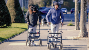
There is further indications that human cartilage can regenerate. The so -called joint distribution is currently being tested as a therapy option for patients with far advanced knee arthrosis who are too young for a prosthesis. (Knee prostheses last 15 to 20 years, after which they have to be replaced in a complicated operation.) In the process, external fixators are created above and below the knee for six weeks, which look like a kind of screw clamp. They are supposed to push the head and lower leg bones apart by about five millimeters. This opens the joint gap. The patients should continue, but the apparatus reduces the load, so that the knee is supplied with nutrient -containing liquid without being overloaded.
Dutch researchers showed in 2013 that the procedure leads to a slight increase in cartilage and reduces pain – benefits that lasted at least two, in some patients even ten years. Larger clinical trials need to be done on this technique, "but it's a fascinating model," says rheumatologist Philip Conaghan of the University of Leeds in England.
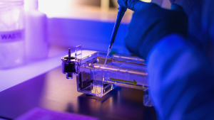
Conaghan is researching new drugs for osteoarthritis. A growth factor called sprifermin appears to slow cartilage loss in some patients. The anti-inflammatory drug canakinumab was originally developed as a cardiovascular drug, but showed a surprising side effect in clinical studies: the recipients required joint replacement much less frequently than a placebo group. Nevertheless, Conaghan warns against too high expectations. Because the repair processes are very slow and difficult to understand, the search for drugs that thicken the cartilage is difficult. "The change is so small that it is difficult to detect it even with the best imaging techniques.«
For the moment, strength training remains the best strategy to strengthen the joints. Conaghan also recommends running in the water: aquajogging. "Strong thigh muscles significantly reduce knee pain - no matter what is going on," he says. "They need strong muscles for everything in life."
© 📓 🐠 LimitedScientific 🇺🇸 , " 👆 💪 5️⃣ 📆 💪 ♻ 🚮 🤢 💪 ⏮ ℹ ⚪️ ➡️ 💉 ⚖️ 😷 ", 2⃣ 0⃣ 2⃣ 2⃣